Microtubules In Animal Cell Diagram - A Labeled Diagram of the Animal Cell and its Organelles ... : The nucleus contains all the genetic material in a cell.
Microtubules In Animal Cell Diagram - A Labeled Diagram of the Animal Cell and its Organelles ... : The nucleus contains all the genetic material in a cell.. Animal cell diagram detailing the various organelles though this animal cell diagram is not representative of any one particular type of cell, it provides insight into the primary organelles and the intricate internal structure of. Even though plant and animal cells are eukaryotic and share a few cell organelles, plant cells are quite distinct when compared to animal cells as they perform different functions. Each replicated chromosome comprises two chromatids, both with the same genetic information. Each centriole is a ring of nine groups of fused microtubules. Helping in cell division by allowing separation of chromosomes
Animal cells come in all kinds of shapes and sizes, with their size ranging from a few millimeters to micrometers. They are long hollow, beaded tubular structure of diameter of about 24nm. The plant cell is rectangular and comparatively larger than the animal cell. Each centriole is a ring of nine groups of fused microtubules. Oct 19, 2020 · therefore, not every animal cell has all types of organelles, but in general, animal cells do contain most (if not all) of the following organelles.
C. elegans oocytes & chromosome segregation: microtubule ... from www.cherrybiotech.com In the complete animal cell centrosome, the two centrioles are arranged such that one is perpendicular to the other. Microtubules (and centrioles) are part of the cytoskeleton. While the plant cell resembles rectangular shape and possesses a rigid cell wall. Helping in cell division by allowing separation of chromosomes Animal cell diagram detailing the various organelles though this animal cell diagram is not representative of any one particular type of cell, it provides insight into the primary organelles and the intricate internal structure of. Even though plant and animal cells are eukaryotic and share a few cell organelles, plant cells are quite distinct when compared to animal cells as they perform different functions. The plant cell is rectangular and comparatively larger than the animal cell. The various cell organelles present in an animal cell are clearly marked in the animal cell diagram provided below.
Some of these differences can be clearly understood when the cells.
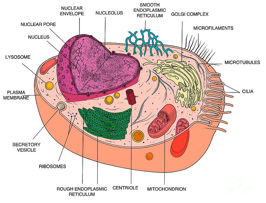
Some of these differences can be clearly understood when the cells. Each replicated chromosome comprises two chromatids, both with the same genetic information. The structure of an animal cell differs slightly from a plant cell, in terms of shape, protective covering and organelles. Microtubules (and centrioles) are part of the cytoskeleton. Oct 19, 2020 · therefore, not every animal cell has all types of organelles, but in general, animal cells do contain most (if not all) of the following organelles. In the complete animal cell centrosome, the two centrioles are arranged such that one is perpendicular to the other. In plant cells, the rigid wall requires that a cell plate be synthesized between the two daughter cells. They are also found in cilia and flagella. They are long hollow, beaded tubular structure of diameter of about 24nm. Helping in cell division by allowing separation of chromosomes In the labeled animal cell diagram, it is nearly circular in shape and lacks outer cell wall; Microtubules are also a part of the cytoskeleton differing from microfilaments in the presence of tubulin protein; And the building blocks of these microtubules are used to grow the mitotic spindle from the region of the centrosomes.
In the complete animal cell centrosome, the two centrioles are arranged such that one is perpendicular to the other. In animal cells, cytokinesis results when a fiber ring composed of a protein called actin around the center of the cell contracts pinching the cell into two daughter cells, each with one nucleus. They are long hollow, beaded tubular structure of diameter of about 24nm. Animal cell diagram detailing the various organelles though this animal cell diagram is not representative of any one particular type of cell, it provides insight into the primary organelles and the intricate internal structure of. Microtubules are also a part of the cytoskeleton differing from microfilaments in the presence of tubulin protein;
Animal Cell Diagram Photograph by Science Source from images.fineartamerica.com Some of these differences can be clearly understood when the cells. They are long hollow, beaded tubular structure of diameter of about 24nm. The nucleus contains all the genetic material in a cell. In plant cells, the rigid wall requires that a cell plate be synthesized between the two daughter cells. In animal cells, cytokinesis results when a fiber ring composed of a protein called actin around the center of the cell contracts pinching the cell into two daughter cells, each with one nucleus. Animal cell diagram detailing the various organelles though this animal cell diagram is not representative of any one particular type of cell, it provides insight into the primary organelles and the intricate internal structure of. The plant cell is rectangular and comparatively larger than the animal cell. And the building blocks of these microtubules are used to grow the mitotic spindle from the region of the centrosomes.
Helping in cell division by allowing separation of chromosomes
Additionally, some organelles will be highly abundant in certain cells and not others. Animal cells come in all kinds of shapes and sizes, with their size ranging from a few millimeters to micrometers. Microtubules are also a part of the cytoskeleton differing from microfilaments in the presence of tubulin protein; In the labeled animal cell diagram, it is nearly circular in shape and lacks outer cell wall; While the plant cell resembles rectangular shape and possesses a rigid cell wall. In plant cells, the rigid wall requires that a cell plate be synthesized between the two daughter cells. Mitosis animation (480 k) or Animal cell diagram detailing the various organelles though this animal cell diagram is not representative of any one particular type of cell, it provides insight into the primary organelles and the intricate internal structure of. Microtubules (and centrioles) are part of the cytoskeleton. In the complete animal cell centrosome, the two centrioles are arranged such that one is perpendicular to the other. The structure of an animal cell differs slightly from a plant cell, in terms of shape, protective covering and organelles. They are also found in cilia and flagella. Each centriole is a ring of nine groups of fused microtubules.
Jul 04, 2020 · animal cell diagram. Jul 27, 2021 · they help in cell division and are involved in the products of various cell surface projections. And the building blocks of these microtubules are used to grow the mitotic spindle from the region of the centrosomes. They are long hollow, beaded tubular structure of diameter of about 24nm. They are also found in cilia and flagella.
Mrs. Remis' Science Blog - 8th grade: CELLS - MICROTUBLES #17 from 4.bp.blogspot.com Each replicated chromosome comprises two chromatids, both with the same genetic information. Centrioles are about 500nm long and 200nm in width that are found close to the nucleus and helps in cell division. Even though plant and animal cells are eukaryotic and share a few cell organelles, plant cells are quite distinct when compared to animal cells as they perform different functions. The nucleus contains all the genetic material in a cell. Microtubules of the cytoskeleton, responsible for cell shape, motility and attachment to other cells during interphase, disassemble. Jul 27, 2021 · they help in cell division and are involved in the products of various cell surface projections. Mitosis animation (480 k) or There are three microtubules in each group.
Microtubules (and centrioles) are part of the cytoskeleton.
Each centriole is a ring of nine groups of fused microtubules. They are long hollow, beaded tubular structure of diameter of about 24nm. Animal cell diagram detailing the various organelles though this animal cell diagram is not representative of any one particular type of cell, it provides insight into the primary organelles and the intricate internal structure of. While the plant cell resembles rectangular shape and possesses a rigid cell wall. Each replicated chromosome comprises two chromatids, both with the same genetic information. In the labeled animal cell diagram, it is nearly circular in shape and lacks outer cell wall; In plant cells, the rigid wall requires that a cell plate be synthesized between the two daughter cells. Microtubules are also a part of the cytoskeleton differing from microfilaments in the presence of tubulin protein; Jul 27, 2021 · they help in cell division and are involved in the products of various cell surface projections. The plant cell is rectangular and comparatively larger than the animal cell. Microtubules of the cytoskeleton, responsible for cell shape, motility and attachment to other cells during interphase, disassemble. There are three microtubules in each group. Microtubules (and centrioles) are part of the cytoskeleton.


0 Komentar